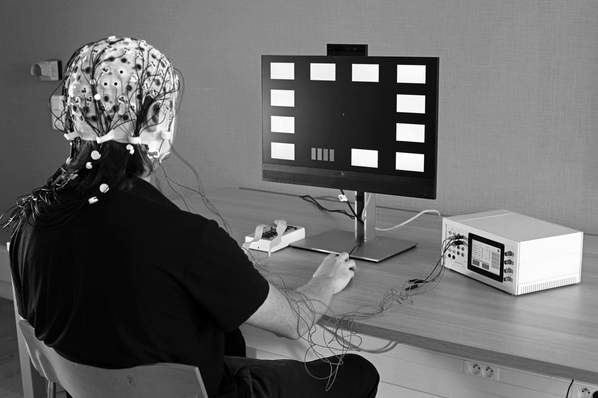
The VBHI is launching a pioneering research project in Europe to evaluate very-low-field brain imaging, with the ambition of making cerebrovascular prevention more accessible to patients, including those living in areas far from major hospital centres.
Identifying the early signs of a disease that can remain silent for a long time
Vascular brain diseases are a major public health challenge. Among them, strokes and vascular-related dementias are major causes of mortality, disability and loss of independence.
To improve their prevention, one key question is now at the heart of research: can damage to the brain’s small blood vessels be detected and measured early enough, before the disease causes symptoms?
This is the aim of the research project led by the VBHI, under the responsibility of Professor Thomas Tourdias, neuroradiologist, Professor and Hospital Practitioner at the University of Bordeaux and Bordeaux University Hospital, and a member of the VBHI management committee.
“The aim is therefore not to replace the high-field MRI used in clinical practice, but to assess which applications could be adequately served by very-low-field MRI, particularly from a prevention and monitoring perspective, bringing these tools closer to the populations concerned,” explains Professor Thomas Tourdias.
Detecting signs of the disease before symptoms appear
Cerebral small vessel disease is extremely common, particularly with advancing age. It can remain silent for a long time while progressively causing lesions that can be seen on brain imaging, known as “markers”.
These markers are associated with an increased risk of subsequent stroke or cognitive impairment. They may also be associated with difficulties with walking and balance, as well as mood disorders.
The aim of the project is to determine whether more readily accessible brain imaging can visualise and quantify these markers sufficiently early and reliably to help delay the onset of symptoms through the prevention strategy developed at the VBHI Institute.
Bringing brain imaging closer to populations
New analysis methods, particularly automated methods, will be developed to identify and measure these markers using very-low-field magnetic imaging. The results will be compared with those obtained using high-field MRI, which is routinely used in clinical care.
One of the advantages of very-low-field imaging lies in the opportunities it offers in terms of compactness and mobility. One of the project’s prospects is to make this imaging approach mobile and deployable outside the hospital, in particular in a mobile medical unit.
The aim would be, if the research results support this approach, to carry out assessments of cerebrovascular health directly within local communities, including in areas far from specialist centres.
Such a development could help improve access to imaging and make it easier to identify people with early markers of cerebral small vessel disease.
The challenge is therefore scientific, medical and territorial: to determine whether a new approach to imaging could contribute to earlier prevention while bringing diagnostic and monitoring tools closer to populations.
A first technological step in Europe
To carry out this research, the VBHI is becoming the first centre in Europe to integrate the second generation of a mobile very-low-field MRI system.
The VBHI is developing its low-field and very-low-field imaging approach through the acquisition of several systems, made possible by €405,033 in funding from the Nouvelle-Aquitaine Regional Council, as well as by government funding awarded to the VBHI under the France 2030 programme.
The project is being conducted within the Biomedical Imaging Platform (pIBio – a CNRS and University of Bordeaux research unit).
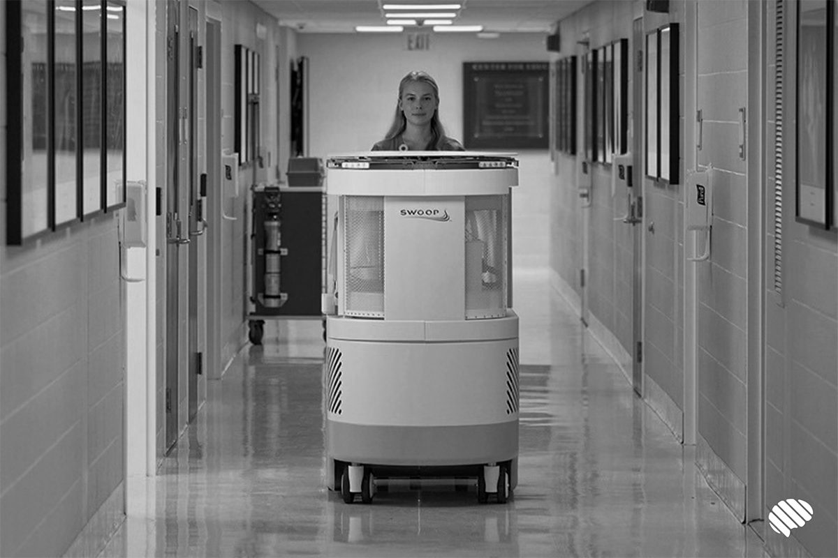
The system used is a Swoop® mobile MRI scanner, developed by Hyperfine and acquired through its French distributor, UpCare.
The Swoop® Portable MR Imaging® system developed by Hyperfine is the first portable magnetic resonance imaging (MRI) system powered by artificial intelligence.
With an ultra-low magnetic field strength of 0.064 Tesla, it enables diagnostic brain imaging to be performed directly at the patient’s bedside, eliminating complex and potentially risky transfers.
UpCare, the French partner and distributor for this technology, is supporting its clinical deployment and will be present at the French-speaking Radiology Days (JFR), taking place from 8 to 11 October in Paris, to present the second generation of this mobile MRI system.
If the results of the study are conclusive, they could pave the way for new approaches to the prevention and monitoring of cerebrovascular diseases, with the prospect of making these approaches accessible to a greater number of people, regardless of where they live.
More info: press release
EM