
CNRS Research Director at LaBRI (Bordeaux Computer Science Research Laboratory) and member of the VBHI management committee, Pierrick Coupé and his team of researchers have just published a study on a new method based on brain imaging and artificial intelligence, called the lifespan tree of brain anatomy, for the simultaneous differential diagnosis of several neurodegenerative diseases. Interview.
What is your research about?
My research field focuses on neuroimaging, particularly on developing new artificial intelligence methods to support radiologists and neurologists.
In this specific study, we developed a new tool based on magnetic resonance imaging (MRI) and artificial intelligence, called the lifespan tree of brain anatomy.
This tool models the evolution of brain structures throughout life (from childhood to old age) by analysing a very large number of subjects.
The idea is to understand how the brain normally changes with age, and how it changes differently in the case of neurodegenerative diseases (such as Alzheimer’s, Parkinson’s, or other forms of dementia).
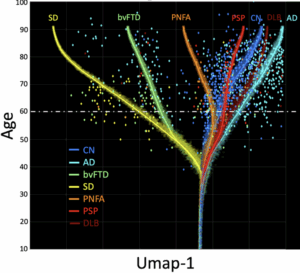
Once the average evolution for each pathology is known as a function of age, this knowledge can be represented in the form of a tree.
A subject can then simply be projected into this space, and by looking at the nearest branches one can identify the potential diseases based on the changes observed on the MRI.
Why focus on the evolution of brain structures to diagnose neurodegenerative diseases?
Neurodegenerative diseases often present with overlapping symptoms (cognitive or motor), which makes clinical diagnosis difficult.
Brain imaging, and particularly the evolution of brain structures across the lifespan, contains potentially useful information for distinguishing between these diseases.
This study introduces a method that models these anatomical changes (volumes of brain structures obtained via MRI) throughout life, to aid in the differential diagnosis of multiple pathologies at once.
Specifically, this new method models changes in the volume of 124 brain structures across the lifespan in order to obtain interpretable representations of brain anatomy during disease progression.
What is differential diagnosis?
In medicine, differential diagnosis consists in determining which specific disease explains a patient’s symptoms, especially when several conditions appear similar. For example, certain memory problems may be linked to Alzheimer’s, but also to other disorders.
The role of differential diagnosis is to avoid confusion and guide patients towards the correct treatment.
In this case, it means distinguishing, for instance, between different forms of dementia or different causes of parkinsonism, to identify which disease is most likely based on brain anatomical signatures.
Why is this a major breakthrough?
At present, the vast majority of available tools only distinguish patients with a specific disease from healthy individuals (binary classification).
Yet for clinicians, the real challenge is not to determine whether a patient is ill or not, but to identify which exact disease they have.
This is precisely where our new tool offers something truly useful: it helps to differentiate between several diseases (multi-class classification), a field still rarely explored in research. This is why we filed a patent for our approach last February.
Moreover, the model is based on the evolution of the brain over the entire lifespan, which provides a richer representation than looking at a single MRI at a given time.
Our tool is therefore not an incomprehensible “black box”: it provides clear, visual representations that are easy for doctors to interpret.
The problem of explainability is one of the main current limitations in the use of AI in medicine. This is what motivated us to choose a readily interpretable representation in the form of a lifespan tree of brain anatomy.
What is the next step?
First, we will make our tool freely available in the coming days on the volBrain platform (www.volbrain.net).
This open-access platform allows brain images to be automatically processed using all the tools developed from our research.
Used by more than 10,000 researchers worldwide, it has already processed over 730,000 MRIs since its creation (volbrain.net/users).
Next, we are working on combining MRI with other medical information (blood tests, cognitive assessments, genetics, etc.) to make the diagnosis even more precise.
Scientific article: Coupé, P., B. Mansencal, J. V. Manjón, et al. 2025. Lifespan Tree of Brain Anatomy: Diagnostic Values for Motor and Cognitive Neurodegenerative Diseases. Human Brain Mapping 46, no. 13: e70336. https://doi.org/10.1002/hbm.70336
EM